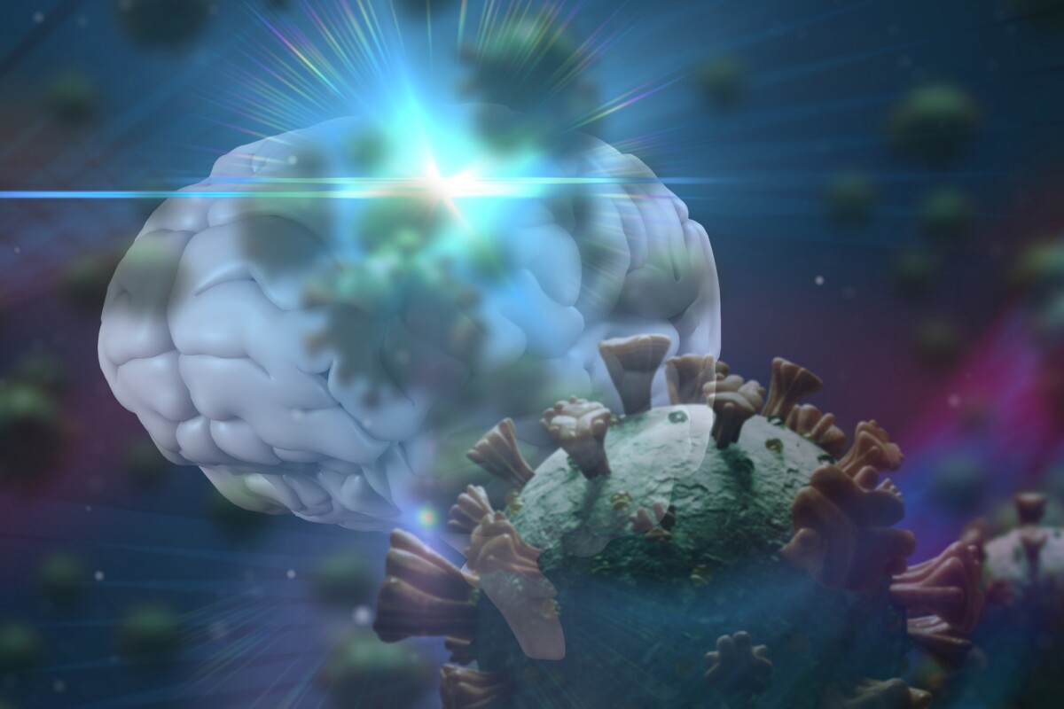
New research led by the Stanford School of Medicine is offering a detailed investigation into brain tissue from those who died from COVID-19. Although no trace of the SARS-CoV-2 virus could be detected, “profound molecular markers of inflammation” were seen, offering clues as to why some patients suffer expansive neurological symptoms following infection.
Stanford neuroscientist Tony Wyss-Coray is perhaps best known for his work transferring plasma from young mice into old mice and discovering it can reverse age-related cognitive decline. Those infamous studies inspired a variety of controversial companies promising to reverse aging through transfusions of young blood.
Wyss-Coray is still investigating exactly what mechanism can explain the mouse findings, and no evidence has yet appeared to show young blood confers those same beneficial effects in humans. One hypothesis is that inflammatory responses in the brain can be triggered by factors in the blood outside of the brain.
As the COVID-19 pandemic took off last year, Wyss-Coray noticed patients frequently reporting neurological symptoms accompanying infections. And many patients still reported neurological symptoms for months after recovering from any acute disease. So two questions framed this new research – can SARS-CoV-2 actually cross the blood-brain barrier to infect a human brain, and what unusual molecular markers can be detected in the brain of a deceased COVID-19 patient?
The research team systematically analyzed 30 brain tissue samples from eight COVID-19 patients and 14 healthy controls. The brain tissue came from the frontal cortex and choroid plexus.
Using single-cell RNA sequencing to measure the expression levels of genes in over 65,000 individual cells, the researchers found a large number of genes linked with inflammatory processes were activated in the brain tissue of COVID-19 patients. Levels of T cells were also more abundant in the COVID-19 patient brains than healthy controls.
“Viral infection appears to trigger inflammatory responses throughout the body that may cause inflammatory signaling across the blood-brain barrier, which in turn could trip off neuroinflammation in the brain,” explains Wyss-Coray. “It’s likely that many COVID-19 patients, especially those reporting or exhibiting neurological problems or those who are hospitalized, have these neuroinflammatory markers we saw in the people we looked at who had died from the disease.”
Wyss-Coray says the molecular markers of inflammation detected in the study share distinct features with what is observed in neurodegenerative diseases such as Alzheimer’s and Parkinson’s.
It is important to note none of the patients displayed any signs of neurological impairment prior to their death. Further work looking for signs of neuroinflammation in cerebrospinal fluid from surviving COVID-19 patients will be necessary to better understand the potential long-term implications.
The study does note there is precedent for an acute viral infection triggering long-term neuroinflammation. So, while the relationship between COVID-19 and neuroinflammation is plausible, it certainly is too early to tell what kind of chronic conditions this all could lead to. Wyss-Coray does hypothesize this potential inflammatory mechanism could explain the symptoms seen in long COVID patients, including fatigue, brain fog and depression.
A more controversial finding in this new study comes from the researchers inability to find any trace of SARS-CoV-2 in the brain tissue of deceased patients. Scientists are still debating whether this novel coronavirus can actually cross the blood-brain barrier and directly infect brain tissue.
A study from Yale University researchers earlier this year found that, hypothetically, the virus can infect brain cells but it is still unknown whether that is actually occurring in real-world infections. Wyss-Coray says his team couldn’t detect any virus in the brain tissue samples they studied.
“We used the same tools they’ve used – as well as other, more definitive ones – and really looked hard for the virus’s presence. And we couldn’t find it,” says Wyss-Coray.
This new research presents the plausible hypothesis that the neurological symptoms associated with COVID-19 and long COVID are due to the way the virus induces peripheral inflammation in the brain, rather than the virus actually directly infecting the brain.
The study concluded by affirming the importance of elucidating these molecular processes as the long-term effects of COVID-19 will not be known for years to come. Other researchers have already warned excessive neuroinflammation triggered by COVID-19 could enhance a person’s risk of developing neurodegenerative diseases such as Parkinson’s, so understanding how this virus can affect the brain will help inform our tracking of the neurological consequences of infection as they emerge.
The new study was published in Nature.
Source: newatlas.com/science/stanford-inflammation-brain-tissue-coronavirus-neurological-long-covid/
